=== Vision disorders
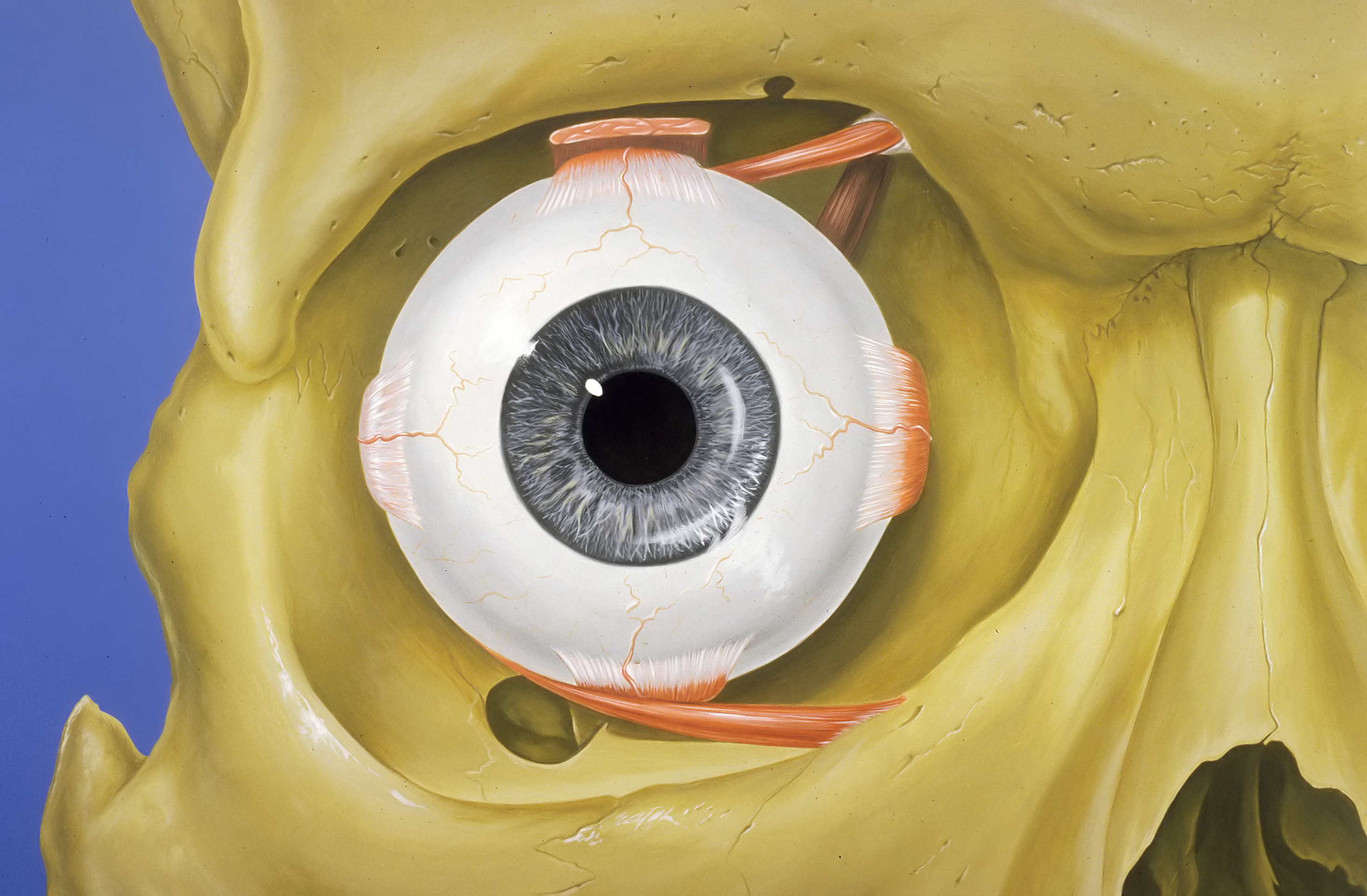
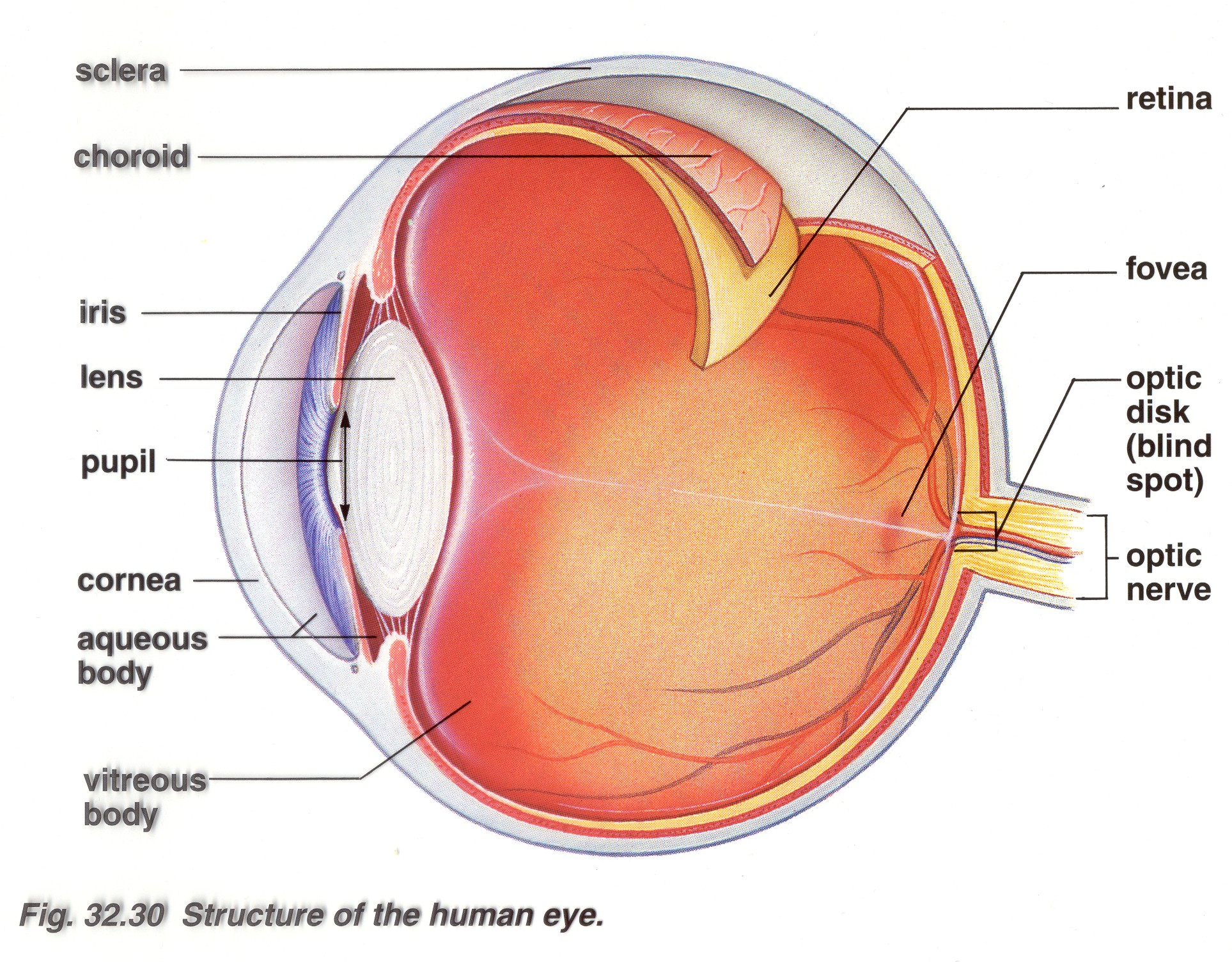
The iris (colored part) of the eye functions like the diaphragm of a camera, controlling the amount of light reaching the retina by automatically adjusting the size of the pupil (aperture). The eye's crystalline lens is located directly behind the pupil and further focuses light rays.

Human eye Wikiwand
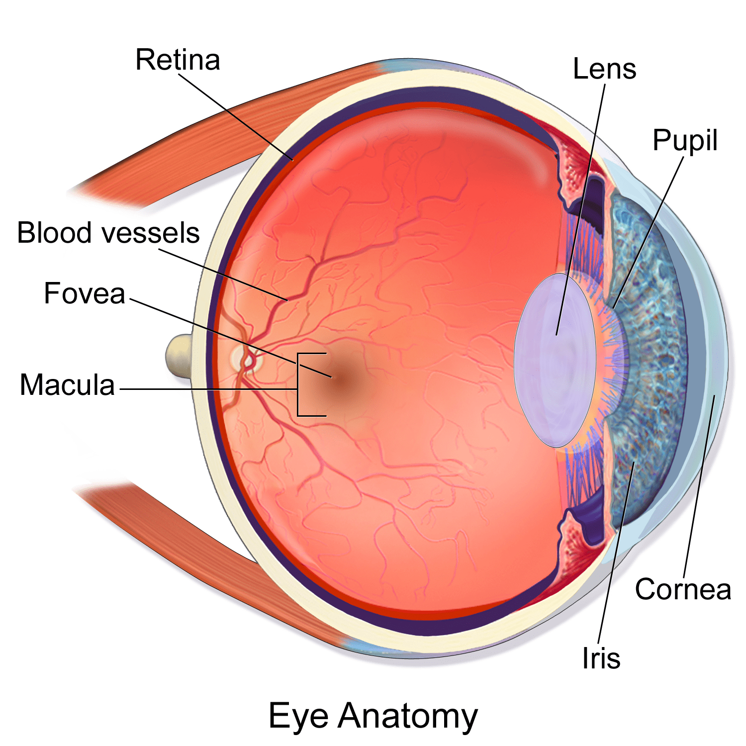
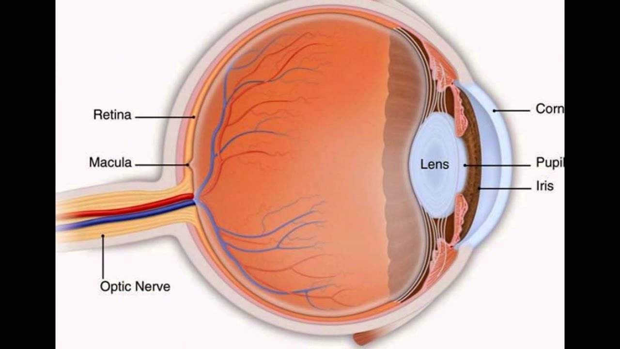
Light projects through your pupil and lens to the back of the eye. The inside lining of the eye is covered by special light-sensing cells that are collectively called the retina. It converts.
Inflammatory Arthritis and Eye Health Prevention, Symptoms, Treatment
Our eye doctors at GHEye excel in prescription of glasses, contact lenses and the diagnosis of a variety of eye diseases. Call our optometrists at (571) 445-3692 to schedule your appointment today for a retinal photo or a standard eye exam. Our eye doctors, Dr. Ally Stoeger and Dr. Jennifer Sun provide the highest quality optometry services and.
20+ Anatomy Of An Eye Pics anatomysystems
Eye Anatomy: The Back of the Eye Dr. Russel Lazarus, October 11, 2020 Did you know that the back of the eye is responsible for transferring visual information from the eye to the brain? In order to see clearly, light is focused onto the back of your eye where it is transformed into electrical impulses.

FileThe human eye.JPG Wikimedia Commons
Retinal detachment describes an emergency situation in which a thin layer of tissue (the retina) at the back of the eye pulls away from its normal position. Retinal detachment separates the retinal cells from the layer of blood vessels that provides oxygen and nourishment to the eye. The longer retinal detachment goes untreated, the greater.

Eye Anatomy
Search from Back Of The Eye stock photos, pictures and royalty-free images from iStock. Find high-quality stock photos that you won't find anywhere else.
ArtStation Eye anatomy photorealistic eyeball Resources
Then they will take pictures of your eye using optical coherence tomography (OCT). With OCT, a machine scans the back of your eye. This provides very detailed pictures of the retina and macula. Your ophthalmologist studies these pictures to check for problems. Macular Hole Treatment. Surgery called vitrectomy is the best way to treat a macular.
FileEye orbit anatomy anterior2.jpg Wikimedia Commons
Mayo Clinic Overview Parts of the eye Enlarge image Retinal diseases vary widely, but most of them cause visual symptoms. Retinal diseases can affect any part of your retina, a thin layer of tissue on the inside back wall of your eye.
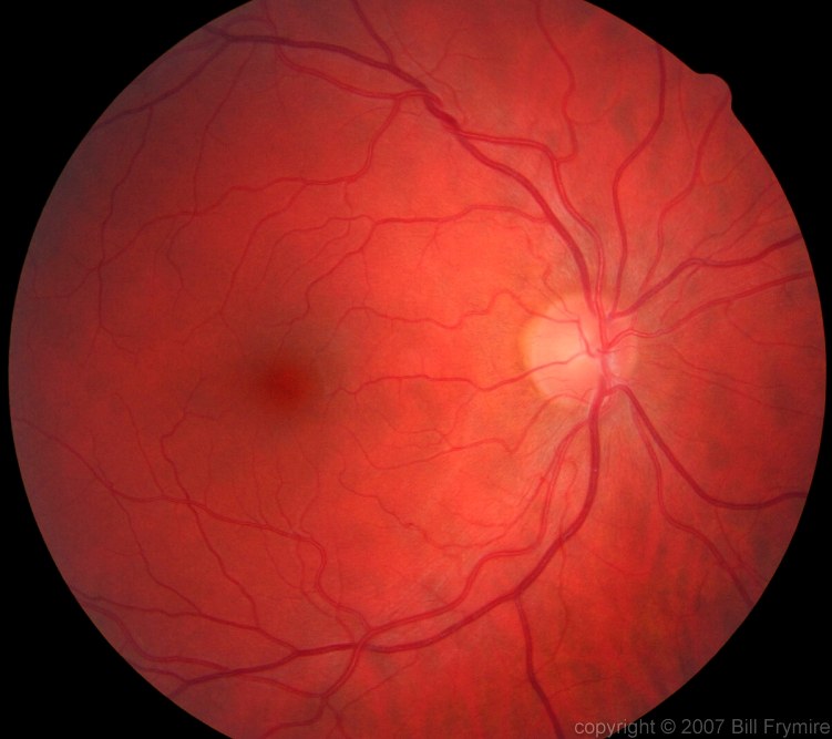
Looking at the back of a retina Image of the Week Bill FrymireBill Frymire
Browse 9,400+ human eye anatomy stock photos and images available, or search for vision or retina to find more great stock photos and pictures. vision retina human eye structure eye chart human eyeball eye doctor eye diagram cataract retinopathy pancreas Sort by: Most popular Anatomy of human eye and descriptions. Components of human eye.
Eye Rolled Back Stock by HaleyOfTheFlame on DeviantArt
Browse 6,269 back of the eye photos and images available, or start a new search to explore more photos and images. NEXT Browse Getty Images' premium collection of high-quality, authentic Back Of The Eye stock photos, royalty-free images, and pictures. Back Of The Eye stock photos are available in a variety of sizes and formats to fit your needs.
This is what human eye looks like from the back. The camera goes through the iris using a laser
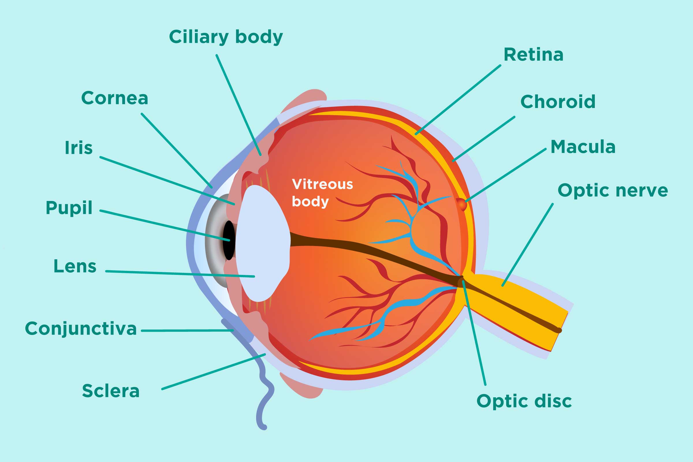
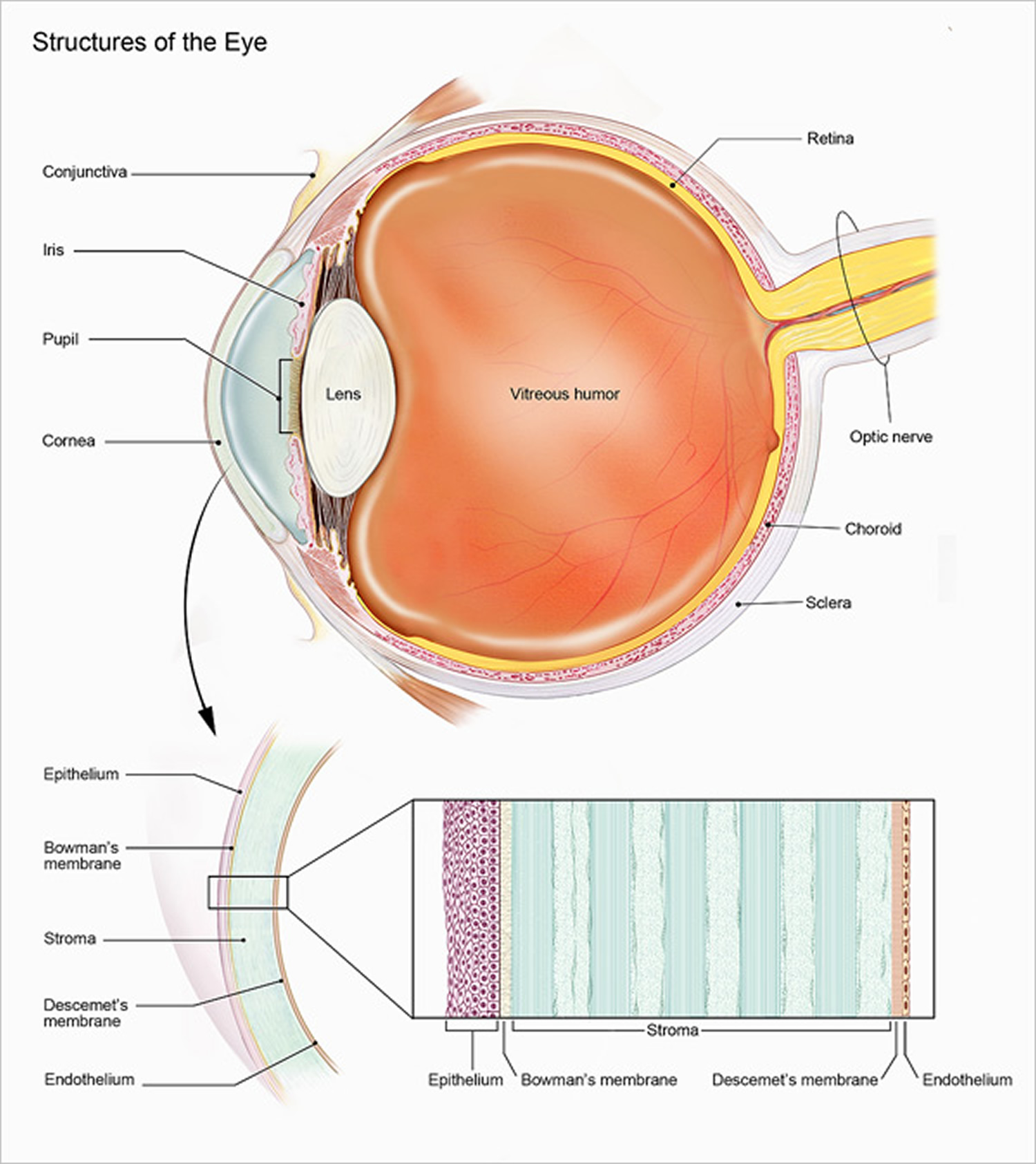
1. Conjunctiva The conjunctiva is the membrane covering the sclera (white portion of your eye). The conjunctiva also covers the interior of your eyelids. Conjunctivitis, often known as pink eye, occurs when this thin membrane becomes inflamed or swollen. Other eye disorders that affect the conjunctiva include:

eye diagram Discovery Eye Foundation
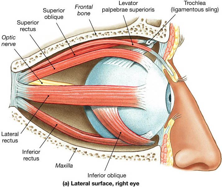
Cornea. The clear, dome-shaped surface that covers the front of the eye. Iris. The colored part of the eye. The iris is partly responsible for regulating the amount of light permitted to enter the eye. Lens (also called crystalline lens). The transparent structure inside the eye that focuses light rays onto the retina. Lower eyelid.
Science of vision How do our eyes enable us to see? How It Works Magazine
Overview What is a retinal hemorrhage? A retinal hemorrhage is the medical term for bleeding in your retina. Hemorrhages are any type of bleeding from a damaged blood vessel. Retinal hemorrhages can be caused by traumas (like getting hit in the head) and health conditions that affect your eyes or blood vessels.
Brain Post How Big is Your Blind Spot? SnowBrains
Muscles in the iris dilate (widen) or constrict (narrow) the pupil to control the amount of light reaching the back of the eye. Directly behind the pupil sits the lens. The lens focuses light toward the back of the eye. The lens changes shape to help the eye focus on objects up close.
The Journey of Light, From the Stars to Your Eyes Universe Today
A choroidal nevus is a freckle located on the back or inner part of your eye. The choroid is part of the uvea, which is the pigmented part of your eye and includes the iris. The choroid falls between the sclera and the cornea. There's such a thing as an amelanotic choroidal nevus, which simply means that the spot is very light in color.
The Lens Of The Eye YouTube
Symptoms. Eye melanoma may not cause signs and symptoms. When they do occur, signs and symptoms of eye melanoma can include: A sensation of flashes or specks of dust in your vision (floaters) A growing dark spot on the iris. A change in the shape of the dark circle (pupil) at the center of your eye. Poor or blurry vision in one eye.